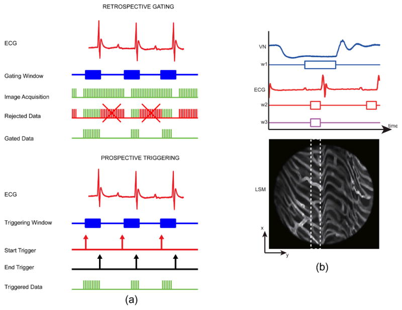
Fig. 4.
(a) Timing diagram of triggering and gating based acquisitions. Retrospective gated acquisition scheme (top): images are continuously acquired while the mouse ECG is simultaneously recorded. Following this non-selective acquisition, only part of the images (patches) that were acquired within the time of a specific gating window are representative of points belonging to the same physical plane. Prospective triggered acquisition scheme (down): images are triggered and acquired only during a specific triggering window which is determined in real time while monitoring cardiac and/or respiratory activity. (b) Double gating of the ECG and the ventilation (VN) signals, and the corresponding minimum artifact area (white dashed line box) within the raw acquired image. The ventilation gating window, w1 is chosen at the time of the end phase of expiration while the ECG gating window w2 is chosen at the time of the end diastole. Both gating windows thus correspond to the time of minimum motion for both lungs and heart, respectively. Reprinted with permission from [14]. © 2012 Landes Bioscience.

