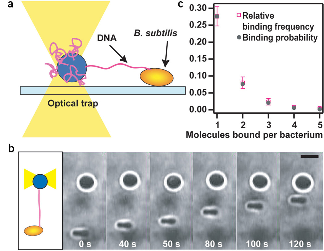
Figure 2.
Experimental setup. (a) One extremity of a DNA molecule was attached to a microscopic bead immobilized by an optical trap. DNA bound to the surface of competent B. subtilis. Deflection of the bead from the center of the laser trap was measured during DNA transport. (b) Time series of a DNA uptake event. As the DNA tether shortened, the distance between the bacterium and the bead decreased. Scale bar, 2 µm. (c) Relative binding frequency and binding probability of DNA binding to one bacterium. ktrap = 0.03–0.05 pN nm−1. n = 250.

