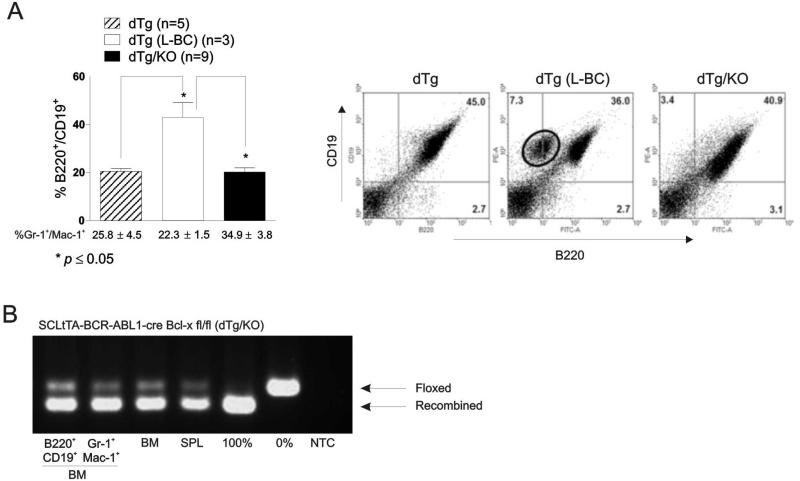
Figure 2. Bcl-x deficient BCR-ABL1 expressing mice do not progress to lymphoid blast crisis.
(A) Left: Flow cytometric analysis shows percentage (mean ± SEM) of B220+/CD19+ and Gr-1+/Mac-1+ cells in the peripheral blood of leukemic dTg, dTg which progressed to lymphoid blast crisis (L-BC), and dTg/KO mice. Right: representative FACS panels of B220/CD19 populations in the spleen of 12 week-induced: dTg (n=5) (left panel), dTg (L-BC; n=3) (middle panel), and dTg/KO (n=12) (far right panel) mice. (B) PCR demonstrates levels of recombination in B220+/CD19+ (lane 1) and Gr-1+/Mac-1+ (lane 2) cells from the marrow, as well as whole bone marrow (BM) (lane 3) and spleen (SPL) (lane 4) of dTg mice 12 weeks after induction. Lane 5 and 6 are controls demonstrating complete and absence of recombination. Results shown are representative of three different mice.

