
Figure 4.
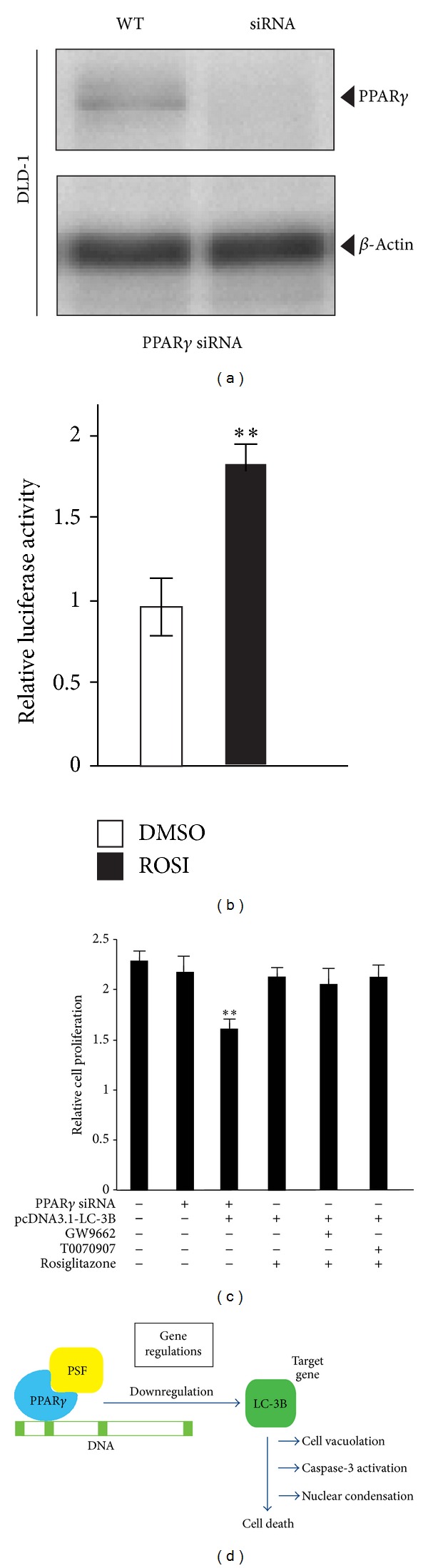
PPARγ activation is not involved in DLD-1 cell proliferation. (a) PPARγ was knocked down in DLD-1 cells. Total protein was extracted from untransfected (WT) and PPARγ siRNA-transfected cells. Forty-eight hours later, whole-cell lysates were subjected to western blot analysis for PPARγ. Incubation with an anti-β-actin antibody was used as a protein-loading control. (b) Effect of rosiglitazone on reporter activation in DLD-1 cells. Cells were transiently transfected with a pGL3-PPRE-acyl-CoA oxidase luciferase reporter vector. The cells were treated with 10 μM rosiglitazone (ROSI) for 20 h. Luciferase activity was normalized to luciferase activity. Data are expressed as mean ± SEM (n = 4; **P < 0.01). (c) PPARγ activation is not involved in PSF-LC3B downregulation in DLD-1 proliferation. Cell growth inhibition was measured using Cell Counting Kit-8 at 72 h after treatment with the vehicle, 10 μM rosiglitazone, 10 μM T0070907, or 10 μM GW9662. Data are expressed as mean ± SEM (n = 3; **P < 0.01). (d) Schematic representation of the proposed mechanism of PSF-LC3B action in DLD-1 cells.