
Figure 4.
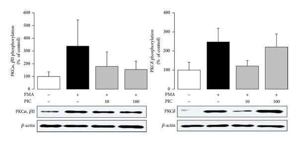
Phosphorylation of PKCα, PKCβII, and PKCδ in PMA-stimulated neutrophils treated with 10 and 100 μmol/L piceatannol (PIC). Phosphorylated PKC isoforms were isolated by Western blotting and detected with anti-phospho-PKCα/βII (Thr638/641) and anti-phospho-PKCδ (Thr505) antibodies. The values are presented as percentage of resting control. Control values, given as optical density of PKC bands corrected to β-actin content, were 78.07 ± 17.86 (PKCα, βII) and 84.84 ± 18.80 (PKCδ). The representative blot manifests elevated phosphorylation of PKC isoforms in neutrophils stimulated with PMA as well as the effect of piceatannol on this increase. Mean ± SEM, n = 4.