Fig. 5.

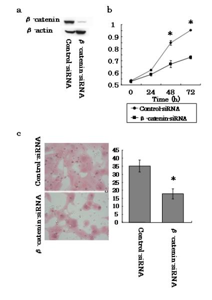
Effects of β-catenin silencing on PanC-1 cells. a Human pancreatic cancer cells were transfected with either control- or β-catenin-siRNA. Total cell lysates were isolated after transfection for 48 h and gal-3 expression levels were detected by western blotting using an anti-human β-catenin antibody. β-actin was used as a loading control. b Proliferation assays. PanC-1 cells transfected with control- or β-catenin-siRNA were cultured in 96-well microplates, and the indicated reagent was injected after 0, 24, 48, or 72 h of culture and the cells were incubated for an additional 2 h. The absorbance was detected using a microplate reader. Data represent the mean ± SD of each treatment (n = 3). c Invasion assays. Cells that invaded through the pores to the lower surface of Matrigel-coated chambers are shown. Invaded cells transfected with control- or β-catenin-siRNA were evaluated on the basis of the mean values from five fields of view at ×200 magnification for each. Data are represented as the mean±SD of each treatment (n= 3). *P < 0.05 versus the control-siRNA.
