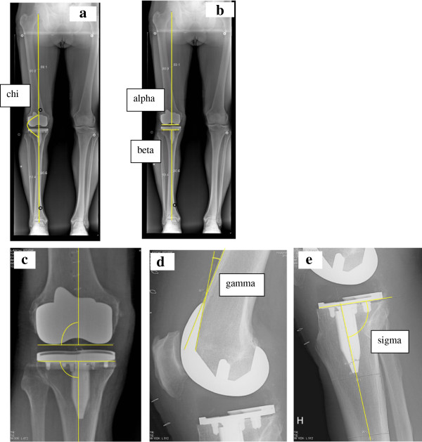
Figure 2.
Radiological measurements. 2a: Drawing tools were used to mark the centre of the femoral head, the knee and talus. Lines connecting these centers define the mechanical axis (chi). The angle is measured on the lateral side. Angles <180° indicate valgus, >180° indicate varus. 2b: Overview of the alpha and beta angles, which measure the femoral and tibial components in the frontal plane. Alpha is measured between a line from the centre of the femoral head to the centre of distal femur and a line parallel to the femoral condyles. Beta is measured between a line from the centre of talus to the centre of proximal tibia and a line along the plateau of tibial component. 2c: The centre of distal femur is defined as the point where a line parallel to the femoral condyles crosses a perpendicular line from the centre of femoral notch. The centre of proximal tibia is defined as the centre of the plateau of the tibial component. 2d: The gamma angle is measured between the frontal femoral cortex and the inner frontal part of the femoral component. A large angle indicates high degree of femoral component flexion. 2e: The tibial slope is measured between the centre of tibia and the plateau of the tibial component, defined as the sigma angle. An angle <90° indicates posterior slope of the tibial component.

