
Figure 2.
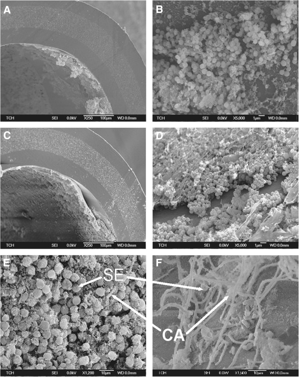
Electron micrographs confirm catheter biofilms in the mouse model of subcutaneous catheter infection. Subcutaneous catheter segments explanted on day 8 of infection were examined by scanning electron microscopy. Electron micrographs of S. epidermidis biofilm infection (A and B) and mixed-species biofilm infection (C, D and E) confirm biofilm formation on catheters in vivo. Mixed species biofilms where predominance of S. epidermidis (Figure 2E) and C. albicans (Figure 2F) are labeled for S. epidermidis (SE) and C. albicans hyphae (CA).