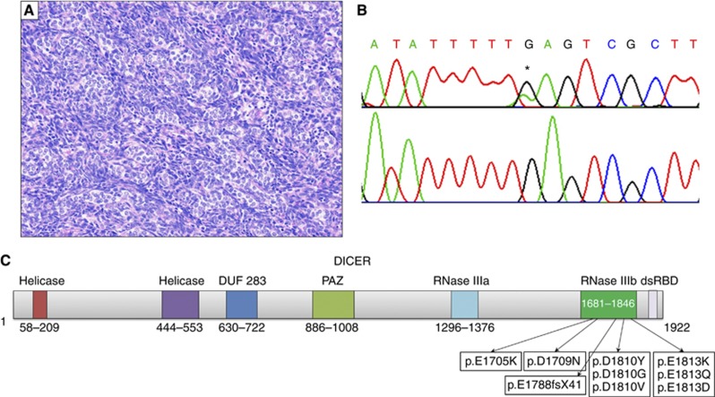
Figure 1.
Diagram of DICER protein and representative SLCT with mutation. (A) Example of poorly differentiated SLCT harbouring a hotspot mutation in DICER1, 20 × . (B) Top: hotspot mutation c.5437G>A found in (A), * denotes mutation. Bottom: wild-type. (C) Schematic of DICER protein (NP_001258211.1), listing all predicted amino-acid changes found in analysed samples. Numbers indicate amino-acid position.

