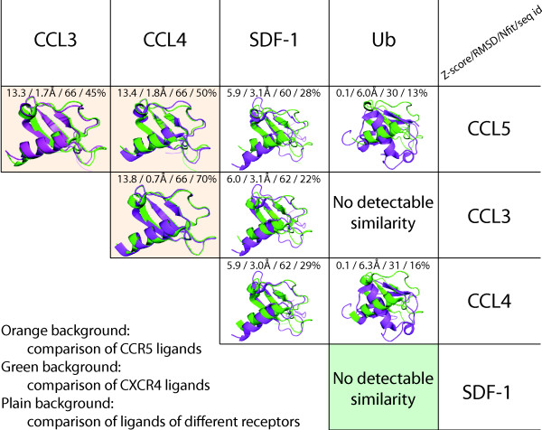
Figure 5.
Structural similarity of endogenous ligands of CCR5 and CXCR4. Each cell presents a structural alignment of two ligands (one shown in green and the other in magenta; for clarity, only alignment of the first chain of the structures of each ligand is shown). The comparison was performed with DaliLite [21], the obtained Z-scores, RMSD (root-mean-square deviation) of Cα atoms, number of aligned residues, and percent of the identical residues in the alignment are reported in each cell.

