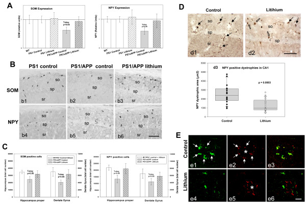
Figure 2.
Lithium treatment avoided neuronal loss in the hippocampus of PS1xAPP mice. (A) The expression of SOM and NPY (n= 20 mice per group) was significantly lower in PS1xAPP control group (ANOVA F(4,96) = 49.63, p = 0.00001. Tukey p < 0.05 or F(4,96) = 27.24, p = 0.0001, Tukey p < 0.05 for SOM and NPY respectively). WT, PS1 (control or lithium) and PS1xAPP lithium displayed no differences. B) SOM (b1-b3) or NPY (b4-b6) immunolabeled sections through CA1 subfield of PS1 control or PS1xAPP control and lithium groups. C) Total SOM or NPY cell number in hippocampus proper and dentate gyrus was assessed by stereology (n = 5 per genotype and treatment). WT and PS1 groups displayed no differences and data were pooled. Only control PS1xAPP displayed a significant reduction in SOM (ANOVA F(2,22) = 14.57, p = 0.00002 or F(2.22) = 11.84, p = 0.0008 for hippocampus proper and dentate gyrus, respectively) and NPY (ANOVA F(2,22) = 17.02, p = 0.0001 or F(2,22) = 6.87, p = 0.008 for hippocampus proper and dentate gyrus, respectively) cell number. D) NPY-positive dystrophies (black arrows) in CA1 subfield of PS1xAPP control (d1) and lithium mice (d2) (open arrows: NPY cell bodies). (d3) Quantitative analysis (n = 4 mice per group; 6 sections per animal) demonstrated the existence of a significant reduction of NPY-positive dystrophic area (μm2) in PS1xAPP lithium. E) Co-localization of SOM (green) and AT8 (red) in dystrophies assessed using confocal microscopy. In PS1xAPP control mice (e1 to e3), SOM positive dystrophies (arrows), surrounding Abeta plaque (asterisk), were positive for AT8. Lithium produced a reduction in both SOM (e4) and AT8 (e5) positive dystrophies and in the co-localization between SOM and AT8 in dystrophies (e6). Scale bars: b1-b6 100 μm; d1 and d2 100 μm; e1-e6 20 μm.

