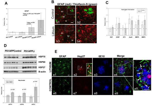
Figure 6.
Lithium increased the astrocyte activation and the Hsps levels. A-B) Lithium treatment increased the astrocyte activation assessed by qPCR (A) and GFAP immunoreactivity (B). GFAP expression (A) was assayed in hippocampal samples from 10 mice per group. GFAP expression increased in PS1xAPP control group and further increased in PS1xAPP lithium-treated mice. ANOVA (F(3,36) = 18.82, p = 0.0001). The post-hoc analysis using Tukey is presented in the graph. B) Representative confocal images through the CA1 field of control (b1, b2) and lithium-treated (b3, b4) mice, showing GFAP-immunoreactive astrocytes (red) and Thioflavin-stained Abeta plaques (green). As shown, GFAP immunoreactivity is clearly higher in lithium-treated mice. C) Lithium did not modify the expression of NGF,GDNF, NT-5, MMP9, MMP3 or ApoE, determined by qPCR (n = 4 mice per group). D) Lithium treatment increased the levels of three Hsps (Hsp70, Hsp60 and Hsp27). Representative western blots and quantitative analysis of Hsps using protein extract from hippocampus of control and lithium-treated PS1xAPP mice (n = 4 mice per group). The expression of all three Hsps was normalized using PS1 control (not shown), and the significance was determined by t-test. E) Confocal images through the CA1 subfield showing GFAP/Hsp27/Abeta triple immunofluorescence labeling. In lithium-treated mice (e1-e5), the core of the Abeta plaques (labeled by 6E10, in blue) is intensely stained with the anti-Hsp27 antibody (in red), as well as astrocytic puncta processes (labeled by GFAP, in green) surrounding the plaques (see insert in e2 showing double GFAP-Hsp27 labeling, and the triple labeling detail in e5). On the contrary, in control mice (e6-e9), the core of the Abeta plaques appears almost devoid of Hsp27 immunostaining and low or no immunoreactivity was observed in astrocytes surrounding the plaques. Scale bars, d1-d9 20 μm, insert in a2 10 μm.

