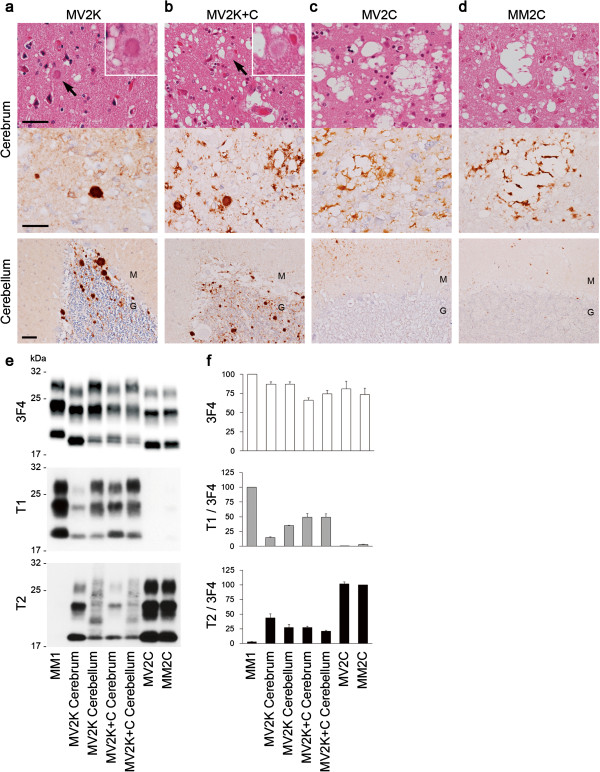
Figure 1.
Histotyping and molecular typing of the MV2 subgroups. (a-d) Histopathological features of MV2K (a), MV2K + C (b), MV2C (c), or MM2C (d) (hematoxylin and eosin stain and immunohistochemistry for PrP). Higher magnification of kuru plaques (arrows) is shown in the insets. M, molecular layer; G, granular cell layer. Scale bar: 100 μm. (e) Western blot analysis of PrPSc in the brains using anti-PrP antibody 3F4 and PrPSc-type specific antibodies T1 or T2. Protein samples were prepared from the frontal cortices. In addition, cerebellar cortices of MV2K or MV2K + C were also examined. (f) The mean signal intensities of 3F4-, T1-, or T2-reactive PrPSc. To compare the amounts of the T1- or T2-reactive PrPSc among the patients, the signal intensities of the T1- or T2-reactive PrPSc were normalized by those of the 3F4-reactive PrPSc. The mean signal intensities of MM1 were assigned as 100 /mm2 in each experiment using 3F4 or T1 antibody, and those of MM2C were assigned as 100 /mm2 in each experiment using T2 antibody. All experiments were repeated independently at least three times.

