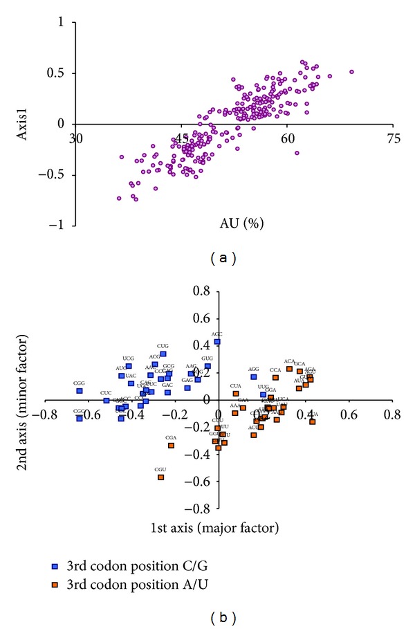
Figure 2.

(a) Correlation between AU content of each retroviral gene and their position on the first axis of CoA. (b) The distribution of synonymous codons is shown along the first and second axes of the CoA. Codons ending with G or C are shown in blue colors, and codons ending with A or U are shown in orange colour.
