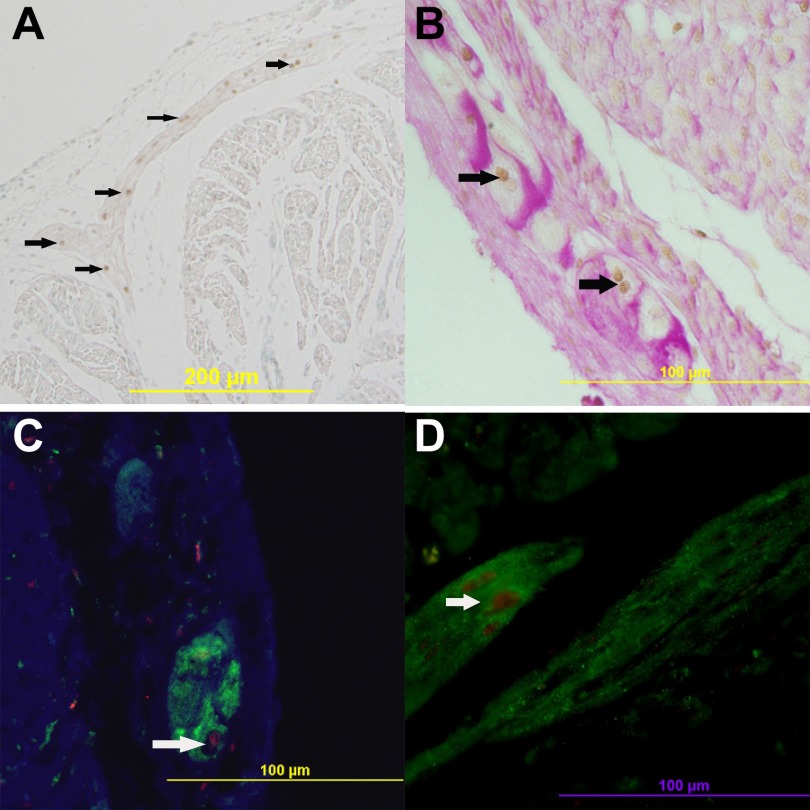
Fig. 3.
Activated caspase-3 nuclei is detected in Purkinje fibers in the fetal heart. A: caspase-3-positive cells (brown nuclei) project into the ventricle wall. B: costaining of periodic and Schiff (PAS) (pink) and caspase-3 (brown nuclei). C: costaining of neurofilament medium (NF-M) (green) and caspase-3 (red). D: costaining of microtubule-associated protein 2 (MAP2) (green) and caspase-3 (red). Arrows show caspase-3-positive Purkinje fibers.

