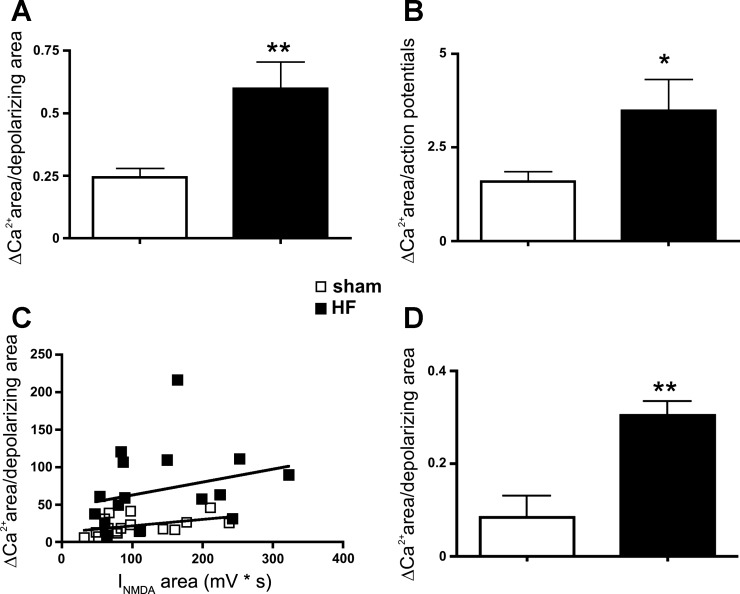
Fig. 3.
Enhanced NMDA-mediated ΔCa2+ per unit of membrane depolarization or evoked action poetential in MNCs of HF rats. Summary data showing a significant increase in NMDA-mediated ΔCa2+ in MNCs from HF rats, when data were normalized either by the total area of the NMDA-mediated depolarization (A) or by the total number of evoked action potentials (B). In C, a plot of the NMDA-evoked ΔCa2+ area as a function of the NMDA-evoked depolarizing area is shown (n = 16 and 18 in sham and HF rats, respectively). D: summary data showing that the larger increase in NMDA-mediated ΔCa2+ per unit of membrane depolarization in HF rats persisted in the presence of tetrodotoxin (1 μM, n = 7 and 4 in sham and HF rats, respectively). *P = 0.05 and **P < 0.01 vs. respective sham.

