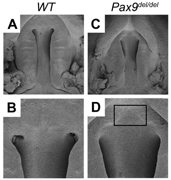
Fig. 3.

Scanning electron microscopy analysis of palate developmental defects in Pax9del/del mutant mice. (A-D) Oral view of the developing palate in wild-type (A,B) and Pax9del/del mutant (C,D) embryos at E13.5. Rectangle in D marks the region of deficiency in primary palate.
