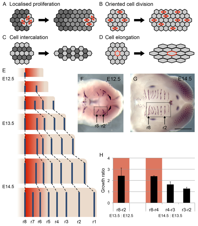
Fig. 1.
Cellular mechanisms of tissue elongation and palatal epithelium development. (A) Localised cell division at the edge of a tissue leads to elongation at that edge. (B) Elongation occurs when daughters of cell divisions are aligned. (C) Tissue elongation through cell intercalation, where cells move perpendicular to the direction of growth, is accompanied by tissue narrowing or thinning. (D) Directional cell elongation can drive tissue elongation. (E) Schematic showing elongation in the palate [right, anterior; top, medial; posterior palate (prospective soft palate) omitted for clarity] using rugae (blue bars) as landmarks for growth. The GZ (red area) is where new rugae are added. (F,G) In situ hybridisation for sonic hedgehog (a marker of developing rugae) showing rugae in developing palate (boxed region) at E12.5 (F) and E14.5 (G) (right, anterior). Positions of ruga 8 and ruga 2 are indicated. Scale bar: 1 mm. (H) Histogram showing ratios of inter-rugal lengths between successive stages, demonstrating elevated growth in the region anterior to ruga 8 (intervals marked in red). Measurements from three samples per stage. Error bars represent s.d.

