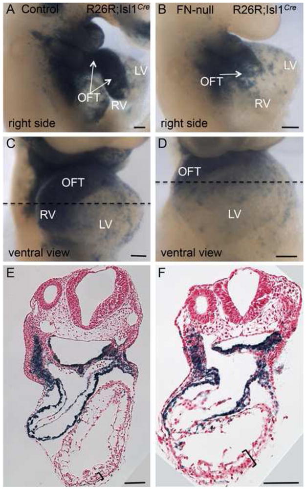
Figure 4. FN is required for the morphogenesis of the cardiac outflow tract and the right ventricle.

Fate mapping of Isl1+ cells and their descendants in control (A, C, E) and FN-null (B, D, F) embryos isolated at E9.5. Note the presence of blue cells (β-gal+) in the outflow tracts of the mutants. Dotted line is the plane of sections shown in E–F. Regions outlined by brackets in E–F show H&E stained sections of thickened myocardial wall in FN-null embryos, they are expanded in Fig. 6.
