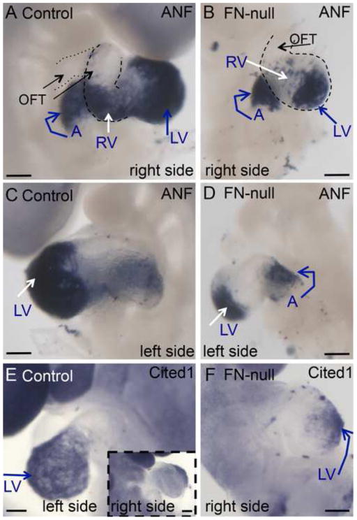
Figure 6. FN is required for normal ventricular morphogenesis.
A–B. Expression of myosin heavy chain is visualized using MF20 antibody. Dotted lines show planes of sections in A′, B′ and B″ below. Blue nuclei show expression of Isl1 protein. Note thickened myocardial wall in the hearts of FN-null embryos (B′, B″, D) compared with controls (A′, C,). E. Quantification of myocardial thickness in serial sections from control and mutant embryos. Ventral ventricular walls in 4–6 sections of serialy-sectioned embryos were measured. In this figure, H&E panels C and D are magnified views of regions indicated by the brackets in Fig. 4E–F.

