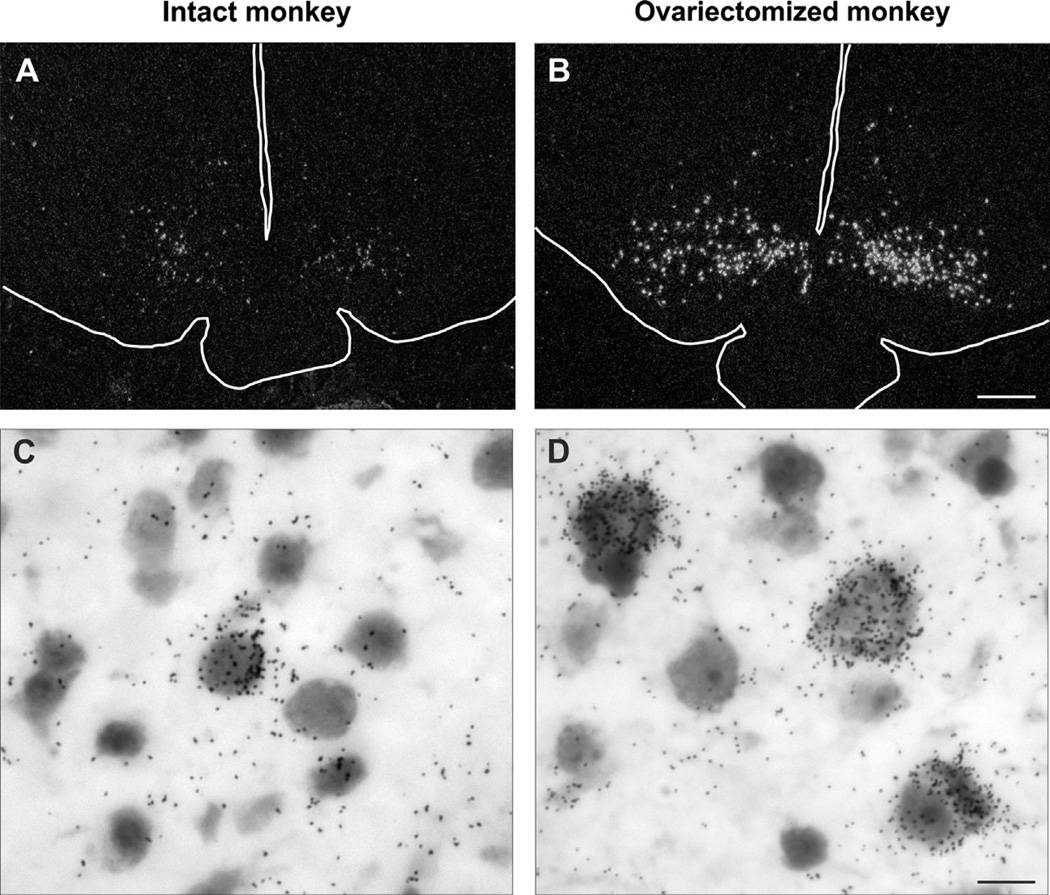
Fig. 2.
(A and B) Darkfield photomicrogaphs of coronal hypothalamic sections from an intact (A) or ovariectomized (B) cynomolgus monkey hybridized with cDNA probes complimentary to NKB mRNA (Tac3 in the monkey) and visualized using autoradiography. The outlines of the base of the brain, pituitary stalk and third ventricle have been drawn on these photomicrographs. Note the marked increase in the number of labeled neurons in arcuate nucleus of the ovariectomized monkey. (C and D) Brightfield photomicrogaphs of neurons expressing NKB mRNA visualized using in situ hybridization in the arcuate nucleus of an intact (C) or ovariectomized (D) cynomolgus monkey. The increase in cell size in the ovariectomized monkeys is accompanied by increased numbers of labeled cells and autoradiographic grains/neuron, indicative of a marked increase in gene expression in response to ovariectomy. These findings mimic the changes in NKB cell size and gene expression observed in the infundibular nucleus of postmenopausal women. Scale bar = 0.5 mm in B (applies to A and B), Scale bar = 25 µm in D (applies to C and D). Modified with permission from Sandoval-Guzmán et al. (2004).

