Figure 4.

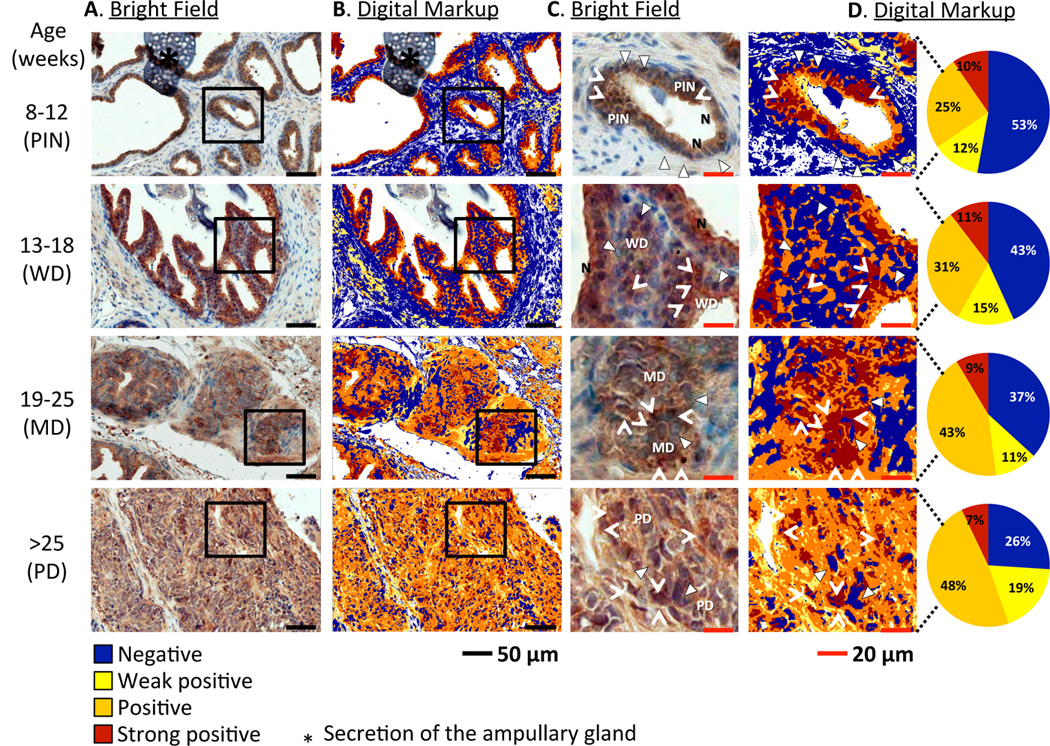
Twist expression in prostate tissues from TRAMP mice varies at different stages of tumor development. A, immunohistochemical analysis of prostate sections from TRAMP mice (n=3/group) of different ages (representing different stages of tumor development). Sections were stained with Twist-DAB and counterstained with hematoxylin. B, digital markup of sections depicted in column A, elaborated by Positive Pixel Count v9 algorithm using Aperio ImageScope image analysis software. C, magnification of tissues within squares in column A. D, magnification of tissues within squares in column and digital quantification of pixels indicating Twist expression: blue = negative, yellow = weak positive, orange = positive, brown = strong positive. Triangles: blue nuclei, negative for Twist. Arrowheads: brown nuclei, positive for Twist. N: normal epithelium. PIN: prostatic intraepithelial neoplasia. WD: well-differentiated adenocarcinoma. MD: moderately-differentiated adenocarcinoma. PD: poorly-differentiated adenocarcinoma. Note absence of Twist expression in nuclei of normal epithelia compared to neoplasias. *: secretion of the ampullary gland.
