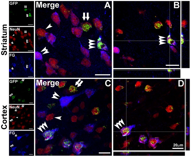
Figure 3. Newborn neurons formed projections to the substantia nigra in ischemic rat brain.
A–D: Confocal microphotographs showed GFP+-NeuN+-FG+ triple-labeling cells (triple arrowheads) in the ipsilateral striatum (A and B) and parietal cortex (C and D) at 13 weeks after MCAO. NeuN+ single-labeling cells, GFP+-NeuN+ and NeuN+-FG+ double-labeling cells were indicated by single arrowhead, double arrows and double arrowheads, respectively. Scale bars are 20 µm.

