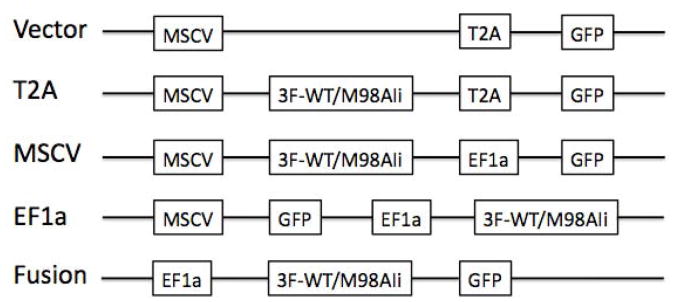
Fig. 1.
Schemes of different pCDH lentiviral vectors. Vectors are named according to the presence or absence of linker (T2A or Fusion) or promoter for Ii in dual-promoter vectors (MSCV or EF1a). Only promoters, transgene (3×Flag tagged Ii) and reporter gene (GFP) are shown. Other elements can be found in the vector maps on System Biosciences website: http://www.systembio.com/lentiviral-technology/expression-vectors/cdna/vector-maps

