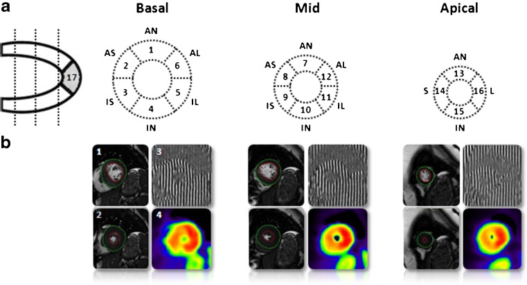
Fig. 2.
The measurement of segmental myocardial efficiency. a. 17-segment model according to the consensus of the American Heart Association. Left ventricular short-axis slices with location of the slices and the names of the segments. AN=anterior, AS=anteroseptum, IS=inferoseptum, IN=inferior, IL=inferolateral, AL=anterolateral, S=septal, L=lateral. b. Left ventricular short-axis cine images in 1. end-diastolic (ED) and 2. end-systolic (ES) phase. 3. CMR tissue tagging image with vertical taglines and 4. Parametric image of [11C]-acetate washout as obtained after kinetic analysis

