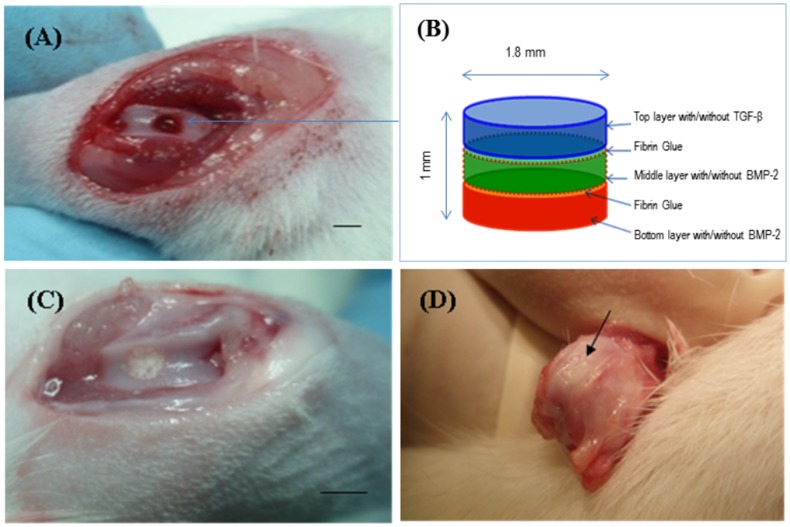
Figure 5. Repair of osteochondral defect in rat patella-femoral groove.
(A) 1.8 mm wide and 1 mm deep osteochondral defects created using a trephine burr. (B) The multi-layered silk fibroin based scaffold with/without growth factors was prepared using 3 scaffold discs by stacking one on top of each other. (C) Scaffolds were implanted to fill the osteochondral defect. (D) After 8 weeks in vivo, the appearance of the repair of osteochondral defect and the interface with surrounding normal cartilage (arrow). Scale bars represented 2 mm.

