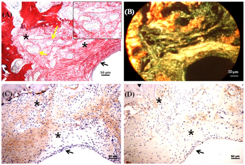
Figure 6. Histological and immunohistochemical appearance of osteochondral defects filled with mulberry silk scaffold (Bm) without growth factors in vivo.
(A) AB/SR staining; (B) Birefringence of AB/SR section; (C) immunostaining with type I collagen and (D) immunostaining with type II collagen. Black asterisks denote the scaffold within the defects. Scale bars represented 50 µm.

