
Figure 6. Erk2 regulates RhoA stability.
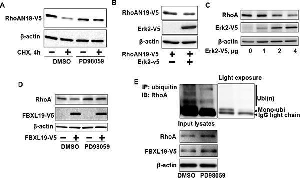
A. RhoAN19-V5 over-expressing MLE12 cells were pretreated with DMSO or PD98059 (10 μM, 1 h) prior to CHX treatment (20 μg/ml, 4 h) and then cell lysates were analyzed for RhoAN19-V5 and β-actin by immunoblotting with V5 tag and β-actin antibodies. B. MLE12 cells were co-transfected with RhoAN19-V5 and Erk2-V5 plasmids and then cell lysates were analyzed for RhoAN19-V5, Erk2-V5, and β-actin by immunoblotting with V5 tag and β-actin antibodies. C. MLE12 cells were transfected with Erk2-V5 plasmids (0 – 4 μg) and then cell lysates were analyzed for RhoA, Erk2-V5, and β-actin by immunoblotting with RhoA, V5 tag, and β-actin antibodies. Shown are representative blots from three independent experiments. D. MLE12 cells were transfected with FBXL19-V5 plasmid and then cells were treated with DMSO or PD98059 (5 μM, 16 h). Cell lysates were analyzed for RhoA, FBXL19-V5, and β-actin by immunoblotting with RhoA, V5 tag, and β-actin antibodies. Shown are representative blots from three independent experiments. E. FBXL19-V5 over-expressing MLE12 cells were pretreated with DMSO or PD98059 (5 μM, 16 h) prior to immunoprecipitation with ubiquitin antibody. The immunoprecipitated complex was analyzed for RhoA by immunoblotting. Input lystes were analyzed for RhoA, FBXL19-V5, and β-actin by immunoblotting with RhoA, V5 tag, and β-actin antibodies. Right panel is a light exposure image.