
Figure 2.
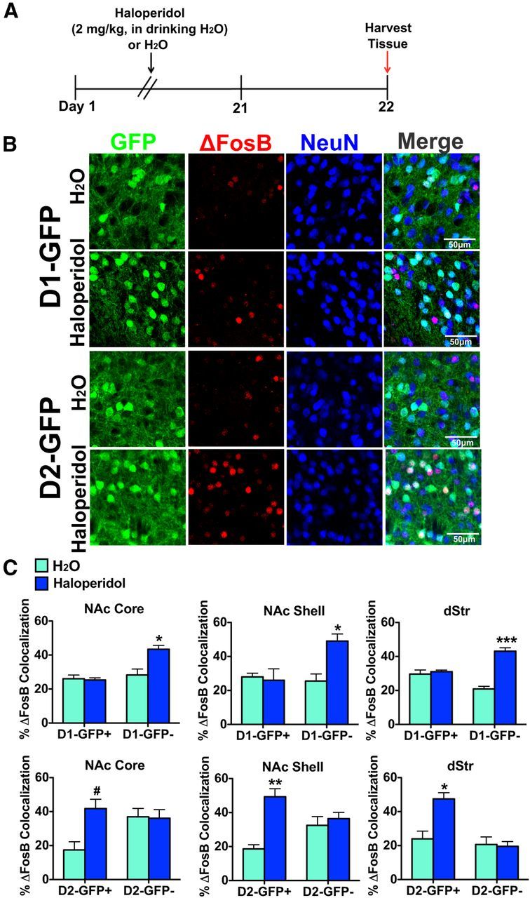
Chronic haloperidol selectively induces ΔFosB in D2-MSNs in striatal regions. A, Time course of 21 d treatment of haloperidol (2 mg/kg, in the drinking water) or water. B, Immunohistochemistry of NAc shell of D1-GFP and D2-GFP mice after haloperidol or water treatment. Immunolabeling for GFP (green), ΔFosB (red), or NeuN (blue) shows induction of ΔFosB in GFP−/NeuN+ neurons in D1-GFP NAc shell or GFP+/NeuN+ neurons in D2-GFP NAc shell. Scale bar, 50 μm. C, Haloperidol treatment significantly induces ΔFosB in GFP−/NeuN+ neurons (D2-MSNs) in D1-GFP mice but not in GFP+/NeuN+ neurons (D1-MSNs) in the same mouse. Two-way ANOVA, NAc core: drug × cell type: F(1,10) = 23.29, p < 0.05, Bonferroni post test: *p < 0.01; NAc shell: drug: drug × cell type: F(1,10) = 30.14, p < 0.05, Bonferroni post test: *p < 0.01; dStr: drug × cell type: F(1,10) = 37.63, p < 0.001, Bonferroni post test: ***p < 0.0001. A significant induction of ΔFosB by haloperidol is also observed in GFP+/NeuN+ (D2-MSNs) but not GFP−/NeuN+ neurons (D1-MSNs) in D2-GFP mice. Two-way ANOVA, NAc core: drug × cell type: F(1,12) = 24.30, p < 0.05, Bonferroni post test: #p < 0.05; NAc shell: drug × cell type: F(1,12) = 26.07, p < 0.01, Bonferroni post test: **p < 0.001; dStr: drug × cell type: F(1,12) = 21.36, p < 0.01, Bonferroni post test: *p < 0.01.