
Figure 7.
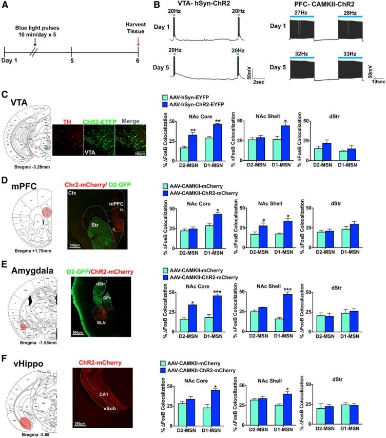
Optogenetic activation of brain regions that innervate the NAc causes distinct patterns of ΔFosB induction in MSN subtypes and striatal regions. A, Optogenetic stimulation paradigm for all conditions. Brains were harvested 24 h after 5 d of optogenetic stimulation. B, Slice physiology recordings of VTA dopamine neurons expressing AAV-hSyn-ChR2-EYFP or mPFC neurons expressing AAV-hSyn-ChR2-mCherry with 1 d of stimulation or 5 d of stimulation. Slices received either phasic 20 Hz, 40 ms blue light (473 nm) pulses (VTA) or 20 Hz blue light pulses (mPFC) C, VTA targeting with AAV-hSyn-ChR2-EYFP or AAV-hSyn-EYFP in D2-GFP mice. We observed an overlap in tyrosine hydroxylase (TH, a marker for dopamine neurons) immunolabeling (red) with ChR2-EYFP expressing neurons (green). Scale bar, 100 μm. Optical stimulation of VTA neurons expressing AAV-hSyn-ChR2-EYFP, for 10 min a day over a 5 d period with mice examined 24 h later, caused a significant induction of ΔFosB in both MSN subtypes in NAc core but only in D1-MSNs in NAc shell. This stimulation parameter caused no induction of ΔFosB in dStr. Two-way ANOVA, NAc core: optogenetic stimuli F(1,16) = 51.97, p < 0.0001, Bonferroni post test: **p < 0.001; (both MSN subtypes) NAc shell: optogenetic stimuli × cell type: F(1,16) = 13.82, p < 0.05, Bonferroni post test: *p < 0.01. D, AAV-CaMKII-ChR2-mCherry or AAV-CaMKII-mCherry as a control was expressed in mPFC of D2-GFP mice. Scale bar, 500 μm. Optical stimulation of mPFC neurons expressing ChR2 for 5 d with mice examined 24 h later resulted in ΔFosB induction in D1-MSNs in NAc core, whereas ΔFosB induction occurred in both MSN subtypes in NAc shell. No induction was seen in dStr. Two-way ANOVA, NAc core: optogenetic stimuli × cell type F(1,14) = 10.31, p < 0.05, Bonferroni post test: *p < 0.01; NAc shell: optogenetic stimuli F(1,14) = 57.17, p < 0.001, Bonferroni post test: #p < 0.05 (D2-MSN), *p < 0.01 (D1-MSN). E, AAV-CaMKII-ChR2-mCherry or AAV-CaMKII-mCherry was expressed in amygdala of D2-GFP mice. Scale bar, 500 μm. Optical stimulation of amygdala neurons expressing ChR2, for 5 d with mice examined 24 h later, induced ΔFosB in both MSN subtypes in the NAc core, and in D1-MSNs in NAc shell, but no change occurred in dStr. Two-way ANOVA, NAc core: optogenetic stimuli F(1,10) = 78.92, p < 0.0001, Bonferroni post test: **p < 0.001 (D2-MSN), ***p < 0.0001 (D1-MSN); NAc shell: optogenetic stimuli × cell type: F(1,10) = 30.31, p < 0.0001, Bonferroni post test: ***p < 0.0001. F, AAV-CaMKII-ChR2-mCherry or AAV-CaMKII-mCherry was expressed in vHippo of D2-GFP mice. Scale bar, 500 μm. Optical stimulation of vHippo neurons expressing ChR2, for 5 d with mice examined 24 h later, caused significant ΔFosB induction in D1-MSNs in both NAc core and NAc shell, with no change observed in dStr. Two-way ANOVA, NAc core: optogenetic stimuli × cell type F(1,10) = 18.30, p < 0.05, Bonferroni post test: *p < 0.01; NAc shell: optogenetic stimuli × cell type: F(1,10) = 22.69, p < 0.05, Bonferroni post test: *p < 0.01.