
Figure 1.
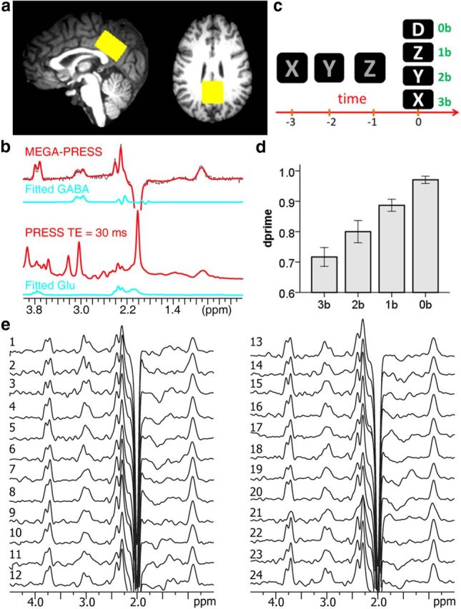
MRS and fMRI experiments. a, A VOI was placed at the PCC/PCu for 1H spectroscopy. b, Regional GABA and glutamate concentrations were assessed using MRS techniques. c, An n-back working memory task was administered to probe the default mode network deactivation, with 0-back as the control condition (time scale is 2 s). d, Behavioral performance (dprime) during the working memory task demonstrated a significant memory load effect. e, Individual GABA spectra. A weighting function of Lorentzian–Gaussian transformation (lb = −3 Hz, gb = 8 Hz) was applied, and the displayed peak intensities were normalized to N-acetylaspartic acid.