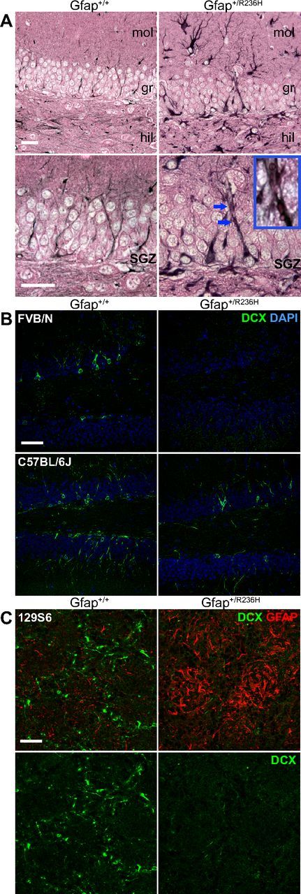
Figure 1.

Hypertrophic radial-glia like cells with Rosenthal fibers and loss of immature neurons in Gfap+/R236H dentate gyrus. A, Immunohistochemical staining for GFAP (black, eosin counter stain) in the granular layer of the dentate gyrus shows intense hypertrophied radial glia-like cells in R236H mutants compared with wild-type mice, which in some cases have eosinophilic inclusion bodies that appear as Rosenthal fibers (bottom row, increased magnification, blue arrows, see inset; n = 4 per genotype, 3 months of age; scale bar, 25 μm; mol, dentate gyrus molecular layer; gr, granular layer; hil, hilus). B, Doublecortin (Dcx) immunofluorescent staining of immature neurons (green) in the dentate gyrus shows almost no staining in GFAP mutant mice compared with wild-type in the FVB/N background and reduced staining in C57BL/6J (n = 3 per genotype, FVB/N = 8 weeks, C57BL/6J = 12 weeks of age; scale bar, 50 μm). C, Dcx immunofluorescent staining (green) in olfactory bulb glomerular layer also shows a reduction of immature neurons in Gfap+/R236H mice with an increase in GFAP expression (red, top only). n = 4 per group, 129S6 at 12 weeks; scale bar, 50 μm.
