Figure 2.

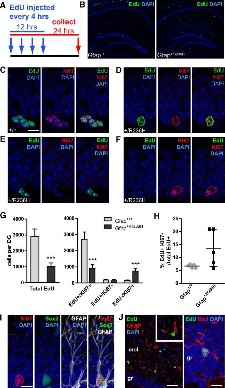
Reduced progenitor proliferation in SGZ of Gfap+/R236H dentate gyrus. A, EdU injection protocol and tissue collection time line for cell proliferation analysis. Arrows indicate injection time points every 4 h for saturated labeling of cycling progenitors followed by tissue collection 24 h after initial injection. B, EdU labeling of dividing cells in the hippocampus of Gfap+/+ mice shows proliferating progenitors in the subgranular zone of the dentate gyrus. In Gfap+/R236H mice, many cells were labeled with EdU outside of the dentate gyrus, with few cells labeled within the SGZ. C–F, EdU and Ki67 labeling in wild-type Gfap+/+ mice (C) shows clusters of dividing cells positive for both markers in the SGZ, whereas EdU+ cells in Gfap+/R236H mice (D–F) are often singular with a different nuclear morphology (D). EdU+/Ki67− cells that have left the cell cycle are apparent in both wild-type and mutant mice (E, Gfap+/R236H shown), and EdU−/Ki67+ cells are frequently apparent in Gfap+/R236H mice (F; scale bar (in C) C–F, 10 μm). G, The total number of cells demonstrating EdU incorporation in the SGZ throughout the rostrocaudal extension of the dentate gyrus (DG) was significantly decreased in Gfap+/R236H mice. The number of cells that were EdU+/Ki67− were similar in both groups, however, Gfap+/R236H mice had many more EdU−/Ki67+ cells than Gfap+/+ mice. Error bars =SD. ***p < 0.0001, two tailed t test between genotypes, n = 5 male mice at 10 weeks of age per genotype. H, The percentage of progenitors leaving the cell cycle (EdU+/Ki67−) was variable in Gfap+/R236H mice, but not significantly increased over that of wild-type mice. I, Immunostaining for Ki67, GFAP, and Sox2 shows colabeling of cells with RGL morphology in the SGZ of Gfap+/R236H mice (FVB/N at 8 weeks, n = 3; scale bar, 10 μm). J, EdU+ cells outside of the SGZ in Gfap+/R236H mice are often GFAP+ astrocytes and sometimes Iba1+ microglia (left scale bar, 50 μm; right scale bar, 10 μm; mol, dentate gyrus molecular layer; gr, granular layer).
