Figure 3.
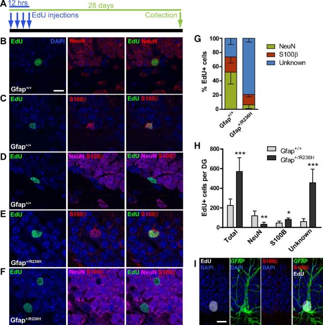
Abnormal differentiation of neural progenitors in Gfap+/R236H hippocampus. A, EdU injection protocol and tissue collection time line for cell fate analysis. Arrows indicate injection time points every 4 h for saturated labeling of cycling progenitors at 10 weeks of age, followed by tissue collection 28 d postinjection. B–D, Analysis of EdU-labeled cell types 4 weeks after injection shows both new neurons (B) and astrocytes (C) in the dentate gyrus of Gfap+/+ mice as shown by costaining with NeuN and S100β, respectively. Unidentified EdU+/NeuN−/S100β− cells were also apparent in Gfap+/+ mice (D). E, F, In Gfap+/R236H mice, this unidentified phenotype accounted for the majority of EdU-labeled cells (F), although many EdU+ cells were identified as S100β+ astrocytes (E). n = 5 male mice at 14 weeks of age per genotype. Scale bar, 10 μm. G, Gfap+/+ mice show a normal distribution of neuronal and astroglial EdU+ cell types in the granular layer of the dentate gyrus (DG), as well as a population of unidentified EdU+ cells at 4 weeks after EdU labeling. Gfap+/R236H mice show a lack of NeuN+ cells with the majority of EdU+ cells negative for either NeuN or S100β. H, The overall number of EdU labeled cells in the granular layer is increased in Gfap+/R236H mice at 4 weeks postlabel compared with Gfap+/+ mice. Although EdU+/NeuN+ neurons are depleted, EdU+/S100β+ astrocytes and especially NeuN−/S100β− cells are increased in mutant mice. *p < 0.05, **p < 0.01, ***p ≤ 0.001, two tailed t test, n = 5 male mice at 14 weeks of age per genotype. I, Immunolabeling shows the presence of EdU+/GFAP+/S100β− cells in the SGZ of Gfap+/R236H mice. Scale bar, 10 μm.

