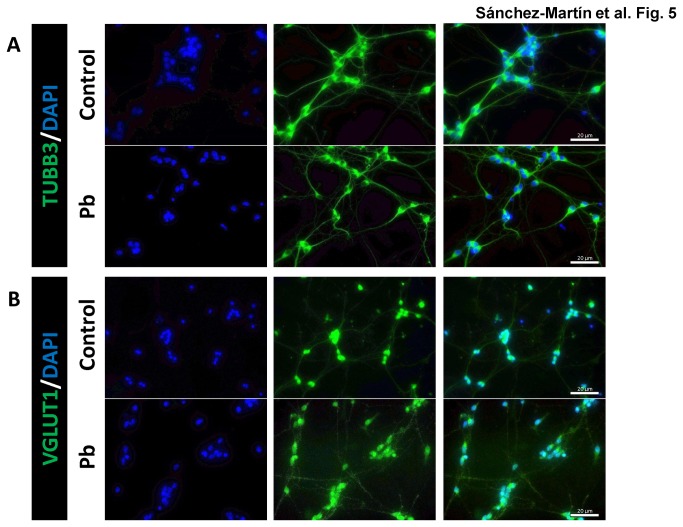
Figure 5. Immunofluorescence detection of TUBB3 (A) and VGLUT1 (B).
Neurons obtained from mESC neural differentiation 3 days after CA disaggregation were untreated (control) or treated with 0.1 µM (Pb) during the whole differentiation process. The third panel shows the merge for each pair of proteins. The experiment shown is representative of three independent cultures.

