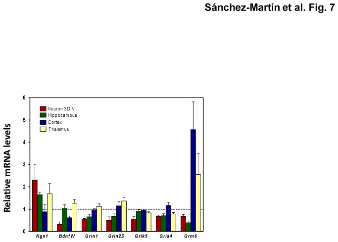
Figure 7. Expression patterns of genes with similar alterations in neural differentiated neurons treated with 0.1 µMPb and in mouse brain gestationally exposed to 3 ppm Pb.
Neurons were treated with 0.1 µM Pb or left untreated for the length of the neural differentiation process. Mouse tissues were the same as in Figure 1. Gene expression was normalized to Gapdh expression and expressed relative to the corresponding levels in untreated or unexposed controls with no Pb treatment. The data shown are the mean ± SEM of three independent determinations.

