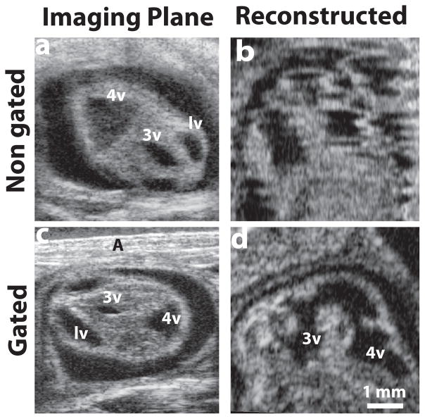
Figure 2. Respiratory-gating to minimize motion artifacts.
(a) A selected plane from the acquired 3D stack without gating, and (b) a reconstructed sagittal plane of the same E12.5 mouse embryo, demonstrating obvious motion artifacts. After implementing respiratory gating, (c) a similar acquired plane of an E12.5 mouse embryo, and (d) the motion-corrected sagittal reconstruction. Labels: A, abdominal wall; lv, lateral ventricle; 3v, third ventricle; 4v, fourth ventricle.

