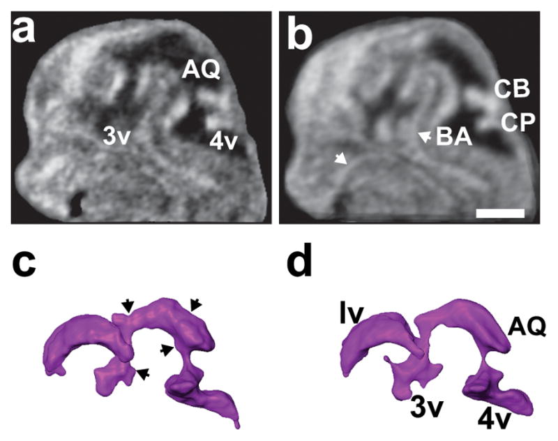
Figure 6. 3D registration and averaging reduces segmentation errors.

Mid-sagittal sections of (a) an individual E12.5 embryo head, and (b) the average of four littermate embryos after voxel-based intensity registration. The registration-averaging significantly smoothed the borders of the ventricles and improved visualization of cerebral blood vessels (white arrows). (c, d) Reconstructions of the brain ventricles in the individual embryo (c) showed a number of surface irregularities (black arrows) that were eliminated by registration-averaging (d). Labels: AQ, aqueduct; BA, basilar artery; CB, cerebellum; CP, choroid plexus; 3v, third ventricle; 4v, fourth ventricle. Scale bar (b) = 1-mm.
