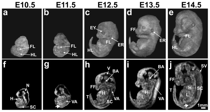
Figure 7. Whole embryo 3D in utero annular array HFU imaging.
Volume reconstructions (a–e) and the corresponding mid-sagittal sections (f–j) from array-focused data for the following embryonic stages: (a, f) E10.5; (b, g) E11.5; (c, h) E12.5; (d, i) E13.5; (e, j) E14.5. Labels: BA, basilar artery; ER, ear; EY, eye; FF, facial features; FL, forelimb; H, heart; HL, hindlimb; L, liver; N, neural tube; SC, spinal cord; SV, spinal vein; T, tail; V, (brain) ventricle; VA, vertebral artery. White arrows (g, j) indicate intersomitic blood vessels. Scale bar (j) = 1-mm.

