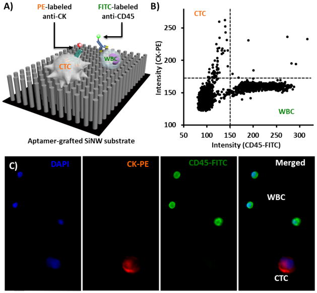
Fig. 3. Immunofluorescence (IF) staining of CTCs immobilized on NanoVelcro substrates.
(A) Schematic representation of an IF protocol developed for identification of CTCs (CK+/CD45−/DAPI+, 40 μm>diameter >10 μm) from non-specifically captured WBCs (CK−/CD45+/DAPI+, 40 μm>diameter >10 μm) and cell debris. (B) An XX-Y scatter plot that summarizes the CK and CD45 expressions of individual cells (including CTCs and WBCs) on a NanoVelcro substrate helps to identify candidate cancer cells. (C) Typical micrographs of a CTC and WBCs immobilized on a NanoVelcro substrate.

