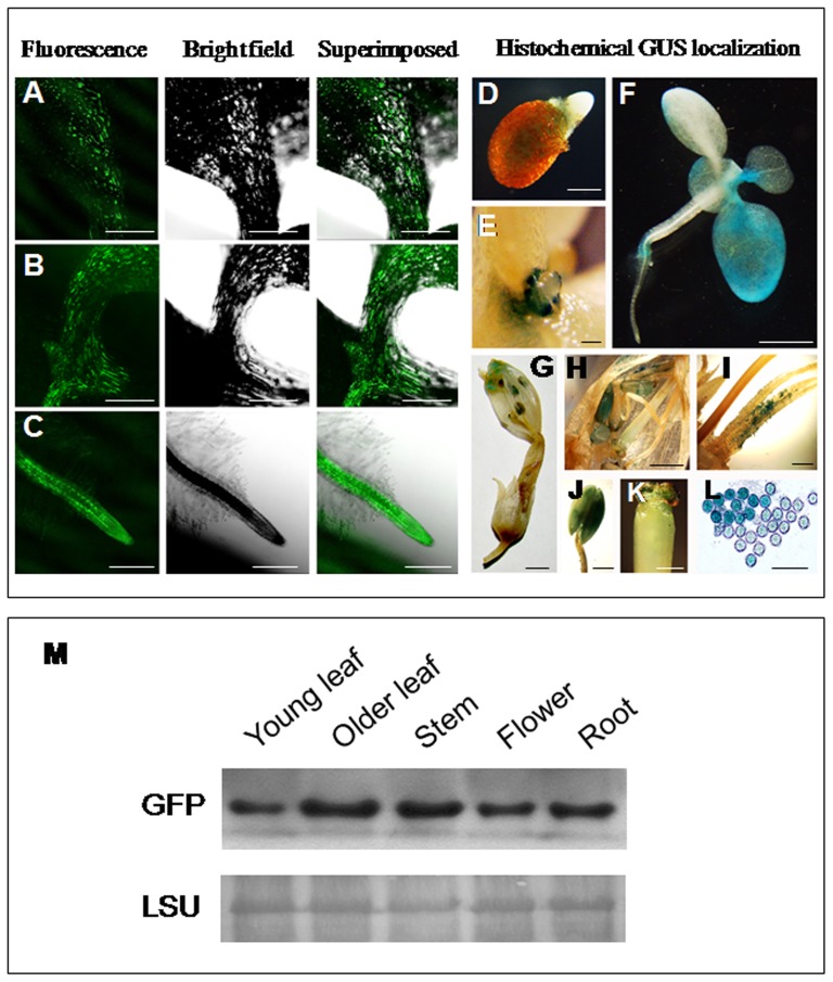
Figure 8. GFP and GUS localization in transgenic tobacco plants generated for the construct GFP::P85–P87::GUS.
(A–C) Confocal laser scanning microscopic analysis of GFP expression under the At4g35985 promoter (P85) in 21-day-old tobacco seedlings (T2 progeny). GFP expression in leaf midrib (A), apical meristematic region (B) and primary root (C) are presented. Green fluorescence image of GFP (left); bright field image (middle), superimposed image (right) are shown. Bar represents 250 µm in each image. Histochemical GUS detection under At4g35987 promoter (P87) in transgenic tobacco plants, GUS expression in germinating tobacco seeds (D) and young primordia at 14-day-old seedlings (E), bar represents 1 mm. (F to H) GUS expression in 21-day-old tobacco seedlings (F); GUS expression in tobacco flower (G); and reproductive tissues of tobacco flower (H); bar represents 5 mm. (I to K) GUS expression in the base of the filament of tobacco flower (I), anther (J) and stigma (K), bar represents 1 mm. (L) GUS expression in pollen grains, bar represents 100 µm. (M) Immunoblot analysis of GFP protein accumulation in various tissue of transgenic tobacco plants generated for the construct GFP::P85–P87::GUS expressing GFP under P85. Western blot showing GFP expression in young leaf, older leaf, stem, flower and root tissues of tobacco plant (T2 generation). Forty µg of total protein from different tissues were subjected to 10% polyacrylamide gel electrophoresis, anti GFP antibody from Santa Cruz Biotechnology (Santa Cruz, CA) was used as primary antibody and horseradish peroxidase-conjugated anti-rabbit secondary antibody (1∶5000) in western blot. Lower panel represents the loading control by detecting Rubisco large subunit (LSU) using Ponceau S stain.

