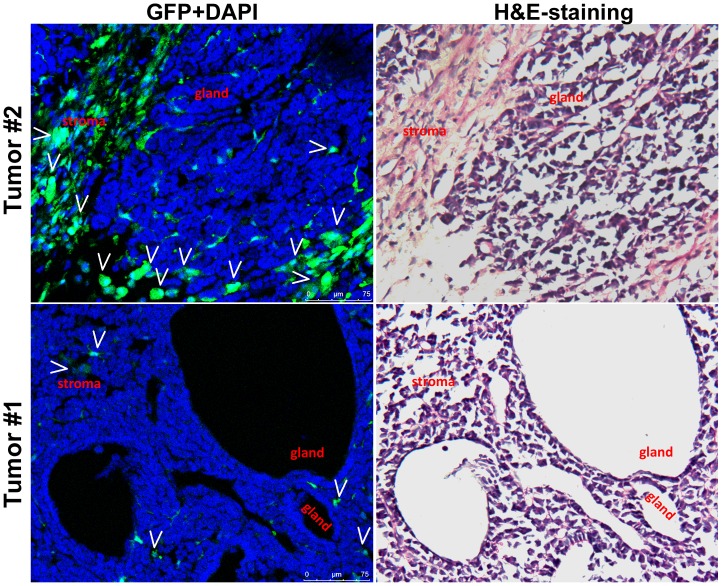
Figure 3. Stromal cells originated from bone marrow-derived cells in two small intestinal tumors.
GFP(+) BMDCs indicated by white arrows were observed in the stromal tissues of two small intestinal tumors in the GFP direct-fluorescence assay (left panel). The histological images were further displayed with the H&E staining (right panel). H&E-staining images of paraffin-embedded tissue from the intestinal tumor used in this direct-GFP confocal analysis are displayed in Figure S3.

