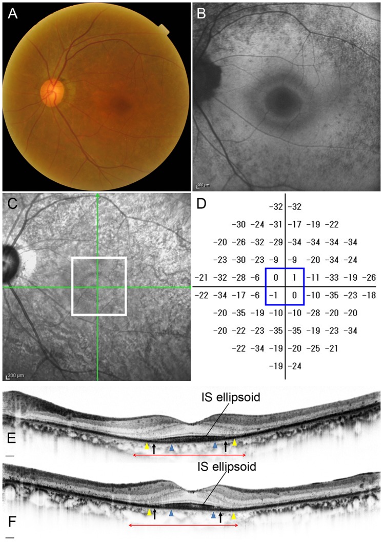
Figure 8. Retinitis Pigmentosa Case (Case 12).
Images of the left eye of a 63-year-old female with RP (Case 12). Snellen equivalent BCVA was 20/15. (A) Fundus photograph shows attenuation of retinal vessels and mottling and granularity of the retinal pigment epithelium. (B) FAF image shows a hyperautofluorescent ring surrounded by a hypoautofluorescent ring in the macula. (C) Infrared image with green arrows indicating the directions of scans shown in E and F, and a white box indicating the area scanned by AO-SLO. (D) Total deviation of Humphrey Field Analyzer (10-2 SITA standard program). Blue box indicates the central 4 points. (E) Horizontal SD-OCT line scan through the fovea. (F) Vertical SD-OCT line scan through the fovea. Blue arrowheads indicate 0.5 mm from the center of the fovea, and yellow arrowheads indicate 1.0 mm from the center of the fovea. The IS ellipsoid is remaining in the area between arrows. Red double-headed arrows indicate the area corresponding to the area scanned by AO-SLO.

