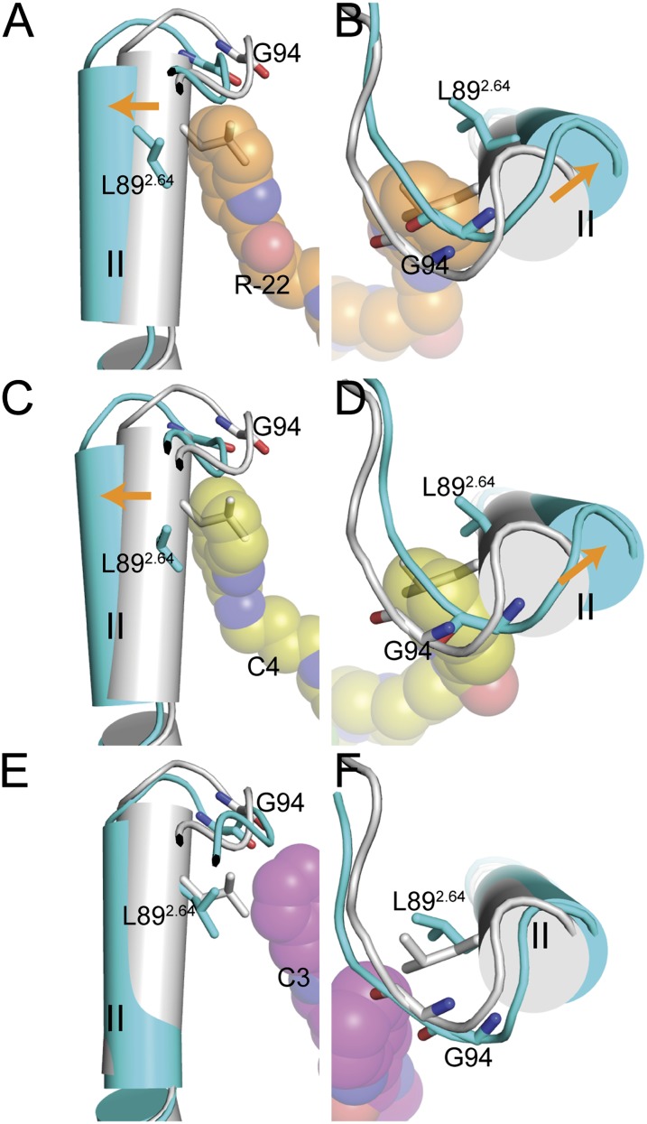
Fig. 4.
Conformational changes induced by R-22 and the C4 and C3 analogs in D3R. Side and top views (left and right, respectively) of the overlaid R-22–, C4-, or C3-bound D3R models (all in cyan) and the eticlopride-bound D3R model (gray) are shown. Conformational changes induced by R-22 (orange spheres in A and B) and C4 (yellow spheres in C and D) in D3R involve the extracellular segment of TM2 (C-terminal to Pro2.59) shifting outward (orange arrows) relative to the eticlopride-bound model, resulting in an approximately 15° larger Prokink wobble angle. In contrast, for the C3 analog (magenta spheres in E and F) the Prokink angles do not differ significantly from the eticlopride-bound model.

