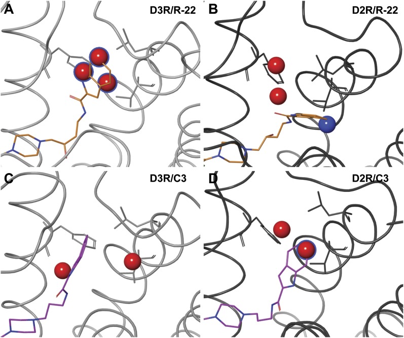
Fig. 5.
Predicted hydration sites in the SBP of D3R (left) and D2R (right) bound to R-22 (orange, top) and C3 (magenta, bottom). High-energy hydration sites with a ΔG >2.5 kcal/mol are also shown as large spheres (Beuming et al., 2009). Hydration sites that are in the Ptm23 subpocket (within 6 Å to the center of mass of Ptm23 residues Val2.61, Leu2.64, and Phe3.28) are colored in red. Sites that are displaced by the ligand (within 2 Å of the indole group) are colored in blue, or have a blue silhouette if they are also in the Ptm23. Multiple high-energy sites are displaced by the aryl amide in the R-22–bound (orange) D3R model (A), but not in the C3 analog–bound D3R model (C), nor the R-22–bound (B) and the C3 analog–bound (D) D2R models.

