Figure 4.

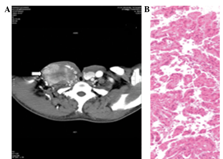
(A) Computed tomography (CT) scan showing a metastatic mass of the right neck. (B) Histopathology showing lymph nodes with metastatic papillary carcinoma (H&E; magnification, ×200).

(A) Computed tomography (CT) scan showing a metastatic mass of the right neck. (B) Histopathology showing lymph nodes with metastatic papillary carcinoma (H&E; magnification, ×200).