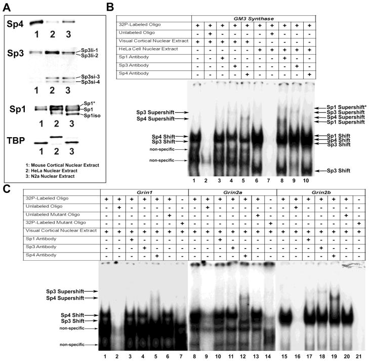
Figure 1.
Relative levels of the Sp factors and In vitro binding of Sp factors to the GM3 Synthase, Grin1, 2a, and 2b promoters. (A) To determine relative levels of Sp factors, equal amounts of nuclear extract from mouse visual cortical tissue, HeLa cell, and N2a cells were probed with Sp4, Sp3, Sp1, or Tata box binding protein (TBP) antibodies. Sp4 levels were highest in mouse cortical tissue, and conversely, Sp3 and Sp1 levels were lowest in mouse cortical tissue as compared to HeLa or N2a cells. In addition, HeLa and N2a cell nuclear extracts contained smaller isoforms of Sp1 (Sp1iso) and Sp3 (Sp3si-3 and Sp3si-4) not present in mouse cortical extract. The phosphorylated form of Sp1 was also found in all nuclear extracts (Sp1*). TBP was used as the loading control and showed similar levels in all nuclear extracts. B and C show In vitro binding of Sp factors to putative binding sites on the GM3 Synthase, Grin1, 2a, 2b, and 2c promoters with EMSA and supershift assays. 32P-labeled oligonucleotides, excess unlabeled oligos as competitors, excess unlabeled mutant Sp oligos as competitors, mouse visual cortical or HeLa nuclear extract, and Sp1, Sp3, or Sp4 antibodies are indicated by a “+” or a “−“ sign. Arrowheads indicate specific Sp1, Sp3, or Sp4 shift, supershift, and non-specific complexes. (B) GM3 Synthase was used as a positive control with mouse cortical nuclear extract (B, lanes 1 – 5) or HeLa cell extract (B, lanes 6 – 10). Incubation with cortical or HeLa nuclear extract revealed specific Sp1, 3, or 4 shift bands (B, lanes 1 and 6, respectively). The location of each band was clearly identified on shorter gel exposure. When excess unlabeled competitor was added, it did not yield a band (B, lanes 2 and 7). The addition of Sp1 antibody yielded two specific supershift bands for HeLa nuclear extract (B, lane 8) corresponding to the presence of tandem Sp binding. The addition of Sp1 antibody did not yield specific supershift bands with cortical nuclear extract (B, lane 3). The addition of Sp3 antibody yielded specific supershift bands with both cortical and HeLa cell extract (B, lanes 4 and 9, respectively), as did the addition of Sp4 antibody (B, lanes 5 and 10, respectively). (C) Incubation of cortical nuclear extract with Grin1, 2a, or 2b probes yielded specific Sp1 and 3 shift bands (C, lanes 1, 8, and 15, respectively). This shift band was competed out with an excess addition of cold probes (C, lanes 2, 9, and 16, respectively). Labeled Grin1, 2a, or 2b probes with mutant Sp sites did not yield specific Sp shift bands (C, lanes 7, 14, and 21). The addition of unlabeled mutant competitor probes to the respective Grin1, 2a, or 2b probes did not compete out the specific shift bands (C, lanes 6, 13, and 20). The addition of Sp1 antibody did not yield supershift bands for Grin1, 2a, or 2b (C, Lanes 3, 10, and 17). The addition of Sp3 antibody did not yield supershift bands for Grin1 or 2a (C, lanes 4 and 11, respectively) but did yield a band for Grin2b (C, lane 18). The addition of Sp4 antibody resulted in supershift bands for Grin1, 2a, and 2b (C, lanes 5, 12, and 19, respectively).

