Fig. 2.
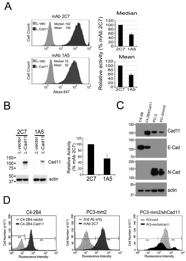
Characterization of mAb 2C7 and mAb 1A5 for their affinity and specificity. (A) Fluorescence activated cell sorting (FACS) analysis showed that binding of mAb 2C7 to L-Cad11 cells was two-fold higher than mAb 1A5. (B) Western blotting analysis comparing the activity of mAb 2C7 and 1A5 showed that binding of mAb 2C7 to Cad11 was two-fold higher than mAb 1A5. (C) Western blotting analysis of cell lysates prepared from C4-2B4, C4-2B4/Cad11, PC3, and PC3-mm2 cell lines for the expression of E-cadherin, N-cadherin, and Cad11. mAb 2C7 does not bind to E-cadherin or N-cadherin. (D) FACS of mAb 2C7 with PCa cell lines. 2C7 shows high fluorescence intensity on C4-2B4-Cad11, but not C4-2B4-vector cells. mAb 2C7 shows high fluorescence intensity with PC3-mm2 cells compared to the cells treated with 2nd antibody only. Knockdown of Cad11 expression by shRNA in PC3-mm2 cells, as described previously (2), significantly decreased the binding of mAb 2C7.

