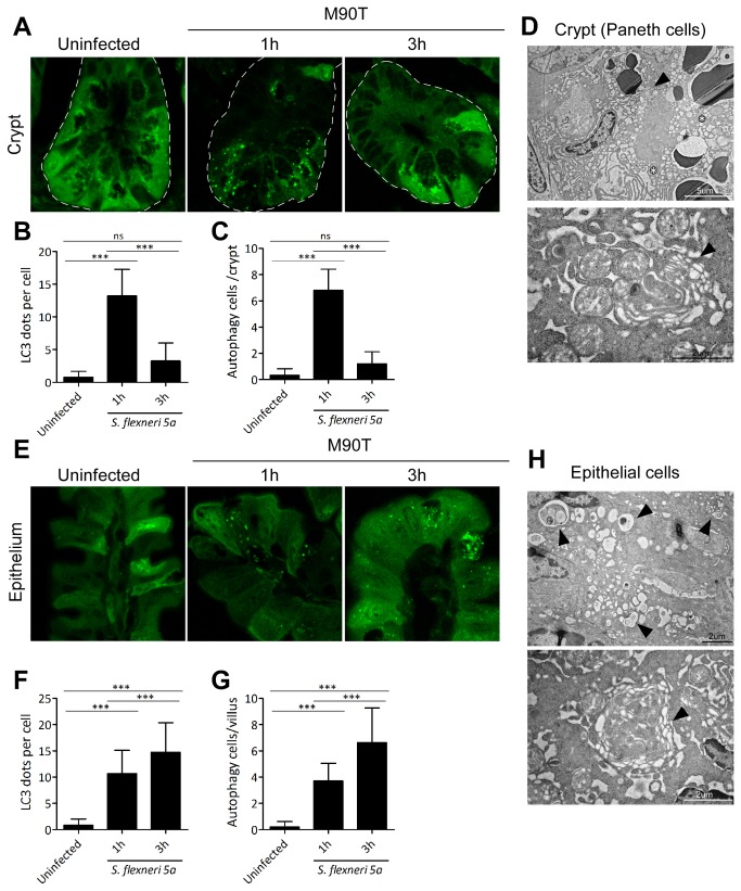
Figure 4. Oral Shigella infection-induced autophagy in Paneth and villous epithelial cells.
After oral M90T infection, crypt (A-C) and villous epithelial cells (E-G) in the terminal ileum region of LC3 GFP transgenic mice were examined for autophagy induction as LC3-GFP puncta formation (n=10). Confocal images of GFP (LC3) puncta formation in the crypt (A) and villi (E). GFP puncta per cell and cells undergoing autophagy in crypt (B-C) and villi (F-G) were counted. TEM images of Paneth cells in the crypt (D) and epithelial cells in the villi (H) of wild type B6 mice 1 h following oral M90T infection. Representative data are from three independent experiments. Multi-layer autophagic vacuole (arrowhead) and excess vacuolization (star). Not significant (ns), ***< P=0.0001.

