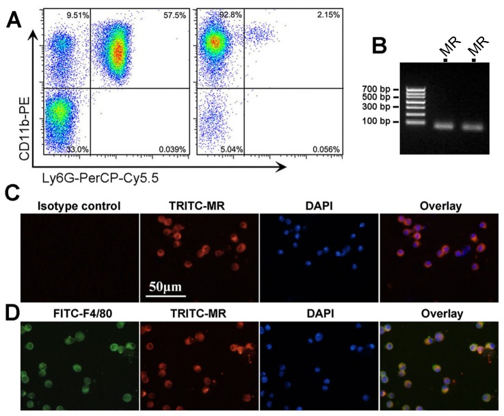
Figure 1. MR is expressed in circulating monocytes and alveolar macrophages.
A shows the purity analysis of enriched Ly6G-CD11b+ monocytes by flow cytometry. The left dot plot shows the monocyte (CD11b+ and Ly6G-) purity is 9.51% before enrichment. After enrichment (the right dot plot), the percent of monocytes is 92.8%. B shows the PCR product agarose gel electrophoresis for MR detection after amplification by real-time PCR (from 2 mice, product length 75 bp). Panel C shows the immunofluorescent staining of purified circulating monocytes. Note that all monocytes are positive for MR (red color). Panel D shows the immunofluorescent staining of cells from mouse BALF. Note that cells positive for F4/80 (green) were also positive for MR (red). Abbreviations: DAPI, 4,6-diamidino-2-phenylindole; FITC, fluorescein isothiocyanate; MR, mineralocorticoid receptor; TRITC, etramethylrhodamine isothiocyanate.

